


Dental Cavity X Ray: Top 5 Surprising Insights 2024
How Dental X-Rays Reveal Cavities
When you hear the word “cavity,” you might picture a visible hole in your tooth. However, many cavities remain hidden from view, lurking in places only detectable by a dental cavity x ray. These X-rays are crucial for identifying tooth decay that the naked eye might miss, particularly in those tight spaces between teeth.
Quick Answer:
– Dental cavity on X-ray: Appears as dark spots in the otherwise white structure of the tooth.
– Importance: Reveals hidden decay, helps in early treatment, prevents severe dental issues.
Cavities Detected on X-Rays:
- Radiolucent: Dark spots indicate holes.
- Radiopaque: Healthy, solid structure appears white.
Understanding how dental cavity x rays work can help you stay ahead of tooth decay and maintain optimal oral health.
As Dr. Ryan Doyle, DDS, with experience in diagnosing dental issues through X-rays, I can attest to the importance of this technology in dental care. By combining advanced imaging with personalized treatment, we ensure your dental health is in the best hands.
What Is a Cavity?
A cavity, also known as tooth decay or dental caries, is a permanently damaged area in the hard surface of your teeth. This damage results in tiny holes or openings that can grow over time if not treated.
How Do Cavities Form?
Cavities form when bacteria in your mouth produce acids that attack the tooth’s enamel, the hard, outer surface of the tooth. Here’s a simple breakdown:
- Bacteria + Sugars/Starches: Bacteria in your mouth feed on sugars and starches from the food and drinks you consume.
- Acid Production: These bacteria produce acids as they digest these sugars.
- Enamel Breakdown: The acids start to break down the enamel, leading to mineral loss.
- Cavity Formation: If the enamel continues to lose minerals and isn’t repaired, it will eventually break down and form a cavity.
Layers of the Tooth
Understanding the layers of a tooth can help explain how cavities develop:
- Enamel: The outermost, hardest part of the tooth.
- Dentin: The layer beneath the enamel, softer and more prone to decay once the enamel is breached.
- Pulp: The innermost part, containing nerves and blood vessels.
Causes of Cavities
Several factors can contribute to cavity formation:
- Poor Oral Hygiene: Not brushing or flossing regularly.
- Sugary Foods and Drinks: Consuming lots of sweets and sodas.
- Lack of Fluoride: Fluoride helps strengthen enamel and prevent decay.
- Dry Mouth: Lack of saliva, which helps wash away food particles and bacteria.
- Frequent Snacking: Constant eating provides a steady supply of sugars for bacteria.
How to Prevent Cavities
Preventing cavities involves a combination of good oral hygiene and healthy habits:
- Brush Your Teeth: Use fluoride toothpaste to brush at least twice a day.
- Floss Daily: Remove food particles between teeth.
- Use Mouthwash: Rinse with an antimicrobial mouthwash to kill bacteria.
- Regular Dental Checkups: Visit your dentist every six months for cleanings and exams.
- Healthy Diet: Limit sugary and acidic foods and drinks.
- Fluoride Treatments: Consider professional fluoride treatments if you’re at high risk for cavities.
By following these steps, you can significantly reduce your risk of developing cavities and maintain a healthy smile.
Next, we’ll delve into How Cavities Form, detailing the process from bacterial activity to enamel breakdown and the role of fluoride in prevention.
How Cavities Form
Cavities begin with a microscopic battle in your mouth. The main culprits? Bacteria, particularly Streptococcus mutans. These bacteria form a sticky film called plaque on your teeth.
The Role of Sugars and Acids
When you eat sugary or starchy foods, the bacteria in plaque feast on them. They produce acids as a byproduct. These acids are the real troublemakers, attacking your tooth enamel.
Enamel Breakdown and Mineral Loss
Your enamel is the hard, outer layer of your teeth. It’s strong but not invincible. The acids from bacteria erode the enamel, causing it to lose essential minerals like calcium and phosphate. This process is called demineralization.
Over time, the enamel weakens and small holes, or cavities, start to form. If left untreated, the decay can reach the softer dentin layer beneath the enamel. Dentin is more vulnerable to acid attacks, so cavities can grow quickly once they reach this stage.
The Role of Fluoride
Fluoride is a mineral that helps repair enamel and reverse early stages of tooth decay. It can be found in toothpaste, mouthwash, and even in some drinking water. Fluoride works by:
- Re-mineralizing Enamel: It helps replace lost minerals, making enamel stronger.
- Inhibiting Bacteria: It reduces the ability of bacteria to produce acids.
- Slowing Enamel Breakdown: It makes enamel more resistant to acid attacks.
Summary
Understanding how cavities form can help you take proactive steps to prevent them. Regular brushing, flossing, and using fluoride products can keep your teeth strong and healthy.
Next, we’ll explore What Does a Cavity Look Like on an X-Ray, providing insights into early detection and the importance of dental X-rays.
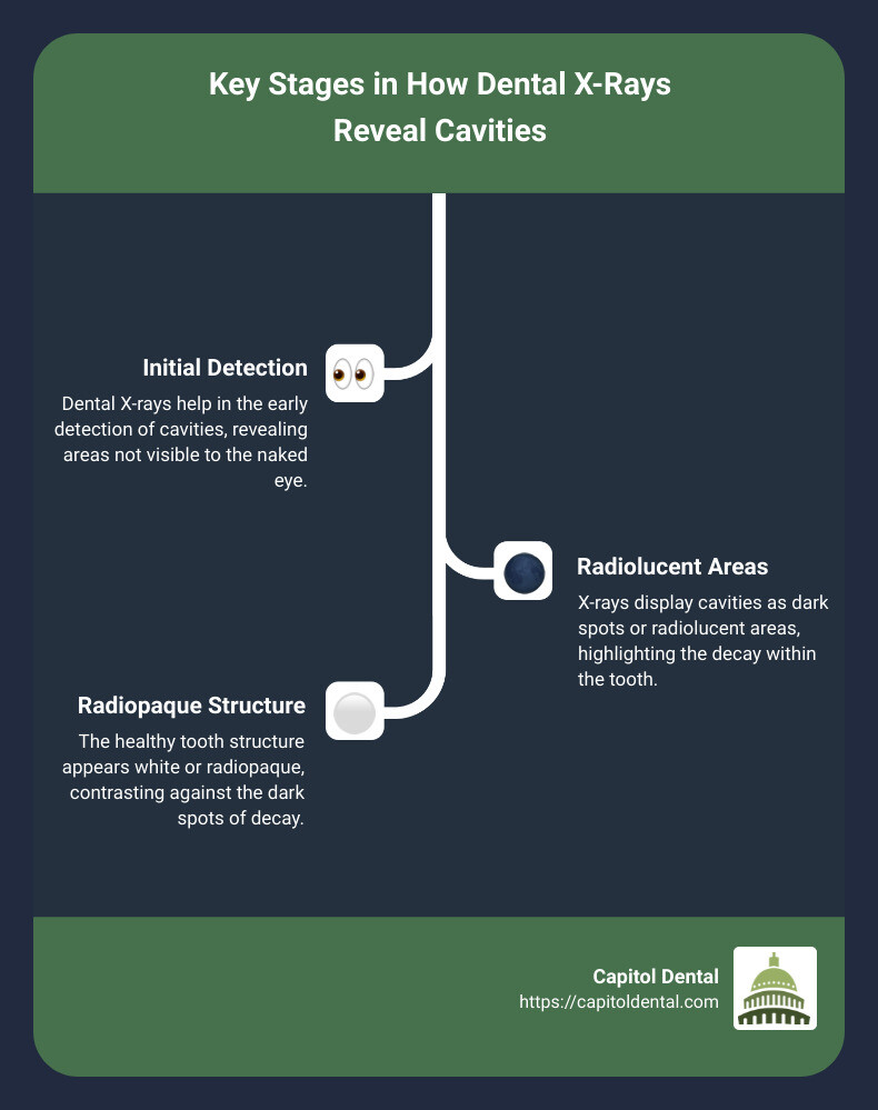
What Does a Cavity Look Like on an X-Ray?
When you look at a dental X-ray, cavities don’t look like they do in your mouth. Instead, they show up as dark spots on the X-ray film. Here’s why:
- Radiopaque: This term means “solid.” On an X-ray, solid structures like teeth and bone appear white.
- Radiolucent: This term means “hollow.” Cavities are holes in the tooth structure, so they appear dark.
The white areas on an X-ray are healthy, solid parts of your teeth, while the dark spots indicate cavities.
Types of Cavities Visible on X-Rays
Cavities can form in different parts of the tooth. Here’s how they look and where you might find them:
1. Chewing Surface
– Description: These cavities form on the top part of the tooth where you chew your food.
– X-Ray Appearance: They show up as dark spots on the top surface of the tooth.
2. Between Teeth
– Description: These are the most difficult to spot without an X-ray because they form in the tight spaces between your teeth.
– X-Ray Appearance: They appear as dark areas between adjacent teeth.
3. Under Fillings
– Description: Called recurrent decay, these cavities form under existing fillings.
– X-Ray Appearance: Look for dark spots beneath the white (radiopaque) filling material.
4. In the Nerve
– Description: Very large cavities can reach the nerve, or pulp, of the tooth.
– X-Ray Appearance: These show up as big dark areas that extend into the center of the tooth.
5. On the Root
– Description: These cavities form on the root surfaces, often below the gum line.
– X-Ray Appearance: They appear as dark spots along the root of the tooth.
6. Under Crowns
– Description: Cavities can also form under dental crowns.
– X-Ray Appearance: Look for dark areas beneath the bright white crown material.
Early Detection
Early detection of cavities is crucial. The sooner a cavity is found, the easier it is to treat. Dental X-rays help dentists spot cavities that might not be visible to the naked eye. This is especially important for catching decay between teeth or under fillings.
By understanding what cavities look like on an X-ray, you can appreciate the importance of regular dental check-ups and X-rays for maintaining your oral health.
Next, we’ll discuss the Signs You May Have a Cavity, helping you identify when it’s time to see your dentist.
Signs You May Have a Cavity
Recognizing the signs of a cavity early can save you a lot of pain and trouble. Here are some common symptoms to watch for:
Tooth Sensitivity
If a tooth feels sensitive to hot or cold drinks, it could be a sign of a cavity. Sensitivity happens because the enamel is wearing away, exposing the softer dentin underneath. If the sensitivity doesn’t go away, it’s time to visit your dentist.
Persistent Toothache
A toothache that won’t go away is another red flag. Pain while eating or drinking, especially something sweet, hot, or cold, could mean you have a cavity. Don’t ignore a persistent toothache; see your dentist as soon as possible.
Discolored Spots on the Tooth
You might notice white, brown, or black spots on your teeth. These spots are often early signs of decay. While you may not always see these spots, your dentist can spot them during a check-up or on an X-ray.
Swollen or Bleeding Gums
Red, swollen, or bleeding gums can also be a sign of a cavity. When decay is near the gum line, it can cause inflammation. If your gums are acting up, it’s a good idea to get them checked out.
Small Holes in the Tooth
Sometimes, you can feel a small hole or crack with your tongue. If you see or feel a hole in your tooth, you likely have a cavity that needs treatment.
Bad Breath That Doesn’t Go Away
Bad breath that persists even after brushing and flossing can be a sign of decay. Bacteria in cavities can produce a foul smell. If you have chronic bad breath, a dental visit is a must.
By being aware of these signs, you can catch cavities early and get the treatment you need to keep your teeth healthy.
Next, we’ll explore How Cavities Are Treated, detailing the steps your dentist will take to fix the problem.
How Cavities Are Treated
Treating a dental cavity is a straightforward process that can usually be completed in about an hour. Here’s a breakdown of what you can expect during a cavity-filling procedure:
Filling Procedure
- Consultation and X-Rays: Your dentist will start by explaining the procedure and taking X-rays to locate the decay. This helps in planning the best course of action.
- Local Anesthetic: To ensure you’re comfortable, a local anesthetic is applied to numb the area around the affected tooth. This numbs your teeth, gums, and surrounding skin, reducing any pain or discomfort.
- Drilling Out the Decay: Once numb, the dentist will use a dental drill to remove the decayed portion of your tooth. This step is crucial to prevent further decay and infection.
- Filling the Cavity: After the decay is removed, the empty space is filled with a material that restores the tooth’s shape and function. There are several types of filling materials available:
Filling Materials
- Composite Resin: This is the most popular choice due to its natural tooth color. Composite fillings bond well to the tooth structure and are ideal for visible teeth.
- Metal Fillings: These include gold and silver amalgam. Metal fillings are very durable and can withstand the pressure of chewing, making them suitable for molars.
- Amalgam Fillings: A mix of metals like silver, mercury, tin, and copper. Amalgam is strong and long-lasting but more noticeable due to its dark color.
- Ceramic Fillings: Made from porcelain, ceramic fillings are tooth-colored and resistant to staining. They are durable but can be more expensive than other options.
Ensuring Longevity
A successful filling keeps future decay out of the treated area. However, fillings can wear down over time and may need replacement. Regular dental check-ups help monitor the condition of your fillings.
By understanding these steps and the materials used, you can be better prepared for your next dental visit. Up next, we’ll discuss How to Prevent Cavities to help you maintain a healthy smile.
How to Prevent Cavities
Preventing cavities is all about good habits and regular care. Here’s a simple guide to keep your teeth healthy and cavity-free.
Brushing
Brush your teeth twice a day with fluoride toothpaste. Make sure to brush for at least two minutes, covering all tooth surfaces. Use a soft-bristled brush and gentle, circular motions to avoid damaging your gums.
Flossing
Floss daily to remove food particles and plaque from between your teeth where your toothbrush can’t reach. This helps prevent cavities from forming in these tight spaces.
Mouthwash
Rinse with mouthwash once or twice a day. Look for a fluoride mouthwash to help strengthen your enamel and protect against decay.
Dental Cleanings
Visit your dentist every six months for professional cleanings and check-ups. These visits help catch early signs of cavities and keep your teeth and gums in top shape.
Avoiding Sugary Drinks
Limit sugary drinks like soda, sports drinks, and fruit juices. These beverages can coat your teeth in sugar, which bacteria turn into acids that erode your enamel.
Healthy Diet
Eat a balanced diet rich in fruits, vegetables, and whole grains. Foods high in calcium and phosphorus, like dairy products, nuts, and leafy greens, help strengthen your teeth.
Fluoride Treatments
Consider fluoride treatments at your dentist’s office. These treatments can provide extra protection against cavities, especially if you’re prone to decay.

By following these simple steps, you can significantly reduce your risk of cavities and enjoy a healthier smile. Next, we’ll explore the Types of Dental X-Rays to understand how they help in maintaining oral health.
Types of Dental X-Rays
Dental X-rays are a crucial tool in maintaining oral health. They allow dentists to see what’s happening beneath the surface of your teeth and gums. Let’s break down the different types of dental X-rays and how each one is used.
Intraoral X-Rays
Intraoral X-rays are taken inside your mouth and are the most common type. They give detailed images of your teeth, bone, and supporting tissues.
- Bitewing X-Rays: These show the upper and lower teeth in one area of your mouth. They’re great for spotting decay between teeth and checking bone density. Bitewings don’t usually show tooth roots.
- Periapical X-Rays: These capture the entire tooth, from crown to root. They help diagnose root infections, fractures, and other issues that go deep into the bone.
- Occlusal X-Rays: These provide a broad view of the bite, showing the entire arch of teeth. They’re useful for identifying issues with tooth development or placement, especially in children.
Extraoral X-Rays
Extraoral X-rays are taken outside your mouth. They give a broader view of your jaw and skull.
- Panoramic X-Rays: These offer a wide view of the entire mouth, jaw, and surrounding structures. They’re useful for assessing tooth development, locating impacted teeth, and evaluating the jaw joint.
- Cephalometric X-Rays: These focus on the side view of the head. They help in planning treatments like braces by analyzing the position of the jaw and teeth.
Advanced Imaging
For more detailed views, dentists may use advanced imaging techniques.
- CT Scans: These provide 3D images of your teeth, jaws, joints, nerves, and sinuses. They are often used before procedures like dental implants to check the height, width, and location of the jawbone.
Comparing Digital and Traditional X-Rays
Digital dental X-rays have several advantages over traditional film X-rays:
- Instant Results: Images appear on the computer screen in seconds.
- Enhanced Image Quality: High-resolution images can be zoomed in on and adjusted for better clarity.
- Reduced Radiation: Digital X-rays use up to 80% less radiation than traditional ones.
- Environmental Impact: No need for chemical processing or film, making digital X-rays a greener choice.
- Electronic Storage: Images can be easily stored and shared electronically.
Digital radiography enhances the precision of diagnosis by providing detailed images that can be manipulated for a closer examination. This makes it easier to detect hidden cavities and other issues that might otherwise go unnoticed.
Understanding the different types of dental X-rays can help you appreciate how these tools contribute to maintaining your oral health. Next, let’s address some frequently asked questions about dental cavity X-rays.
Frequently Asked Questions about Dental Cavity X-Rays
Can you see cavities on an X-ray?
Yes, you can see cavities on an X-ray. Cavities appear as dark shadows or radiolucent areas on the X-ray image. This is because cavities are essentially holes or non-solid structures in the teeth, making them less dense and allowing more X-ray beams to pass through, leading to darker spots on the image.
- Early Detection: X-rays can detect cavities long before they become visible to the naked eye. Early detection allows for quicker treatment, preventing more severe dental issues.
- Between Teeth: Cavities often form in the tight spaces between teeth, which are difficult to inspect visually. X-rays help in spotting these hidden cavities.
How often is it safe to have dental X-rays?
The frequency of dental X-rays depends on your oral health needs. According to the American Dental Association (ADA) guidelines, most people with healthy teeth and gums should have dental X-rays taken once every 24 to 36 months.
- Radiation Safety: Modern digital X-rays expose you to significantly less radiation compared to traditional X-rays. This makes them safer for regular use.
- Individual Needs: If you have gum disease, recurring decay, or other oral health issues, you may need more frequent X-rays. Your dentist will recommend the right schedule based on your specific needs.
Can an X-ray show decay under a filling?
Yes, an X-ray can show decay under a filling. This condition is known as recurrent decay.
- Hidden Decay: X-rays can reveal new decay that has formed under an existing filling. This type of decay is not visible during a regular dental exam.
- Infection Detection: Detecting hidden decay is crucial for preventing infections that can lead to more serious dental problems.
- Severity Assessment: X-rays help assess the severity of the decay, guiding the dentist in deciding the best course of treatment.
Understanding these aspects of dental cavity X-rays can help you appreciate their role in maintaining your oral health.
Conclusion
At Capitol Dental, we are dedicated to providing comprehensive care that addresses all your dental needs. Our team leverages advanced technology to ensure precise diagnostics and effective treatments. This includes the use of digital dental X-rays, which are essential for early detection and management of cavities.
We pride ourselves on offering personalized service tailored to each patient’s unique needs. Whether it’s routine check-ups, cavity fillings, or more complex procedures, our goal is to make your dental experience as comfortable and efficient as possible.
Located in Boise, Idaho, Capitol Dental is committed to improving your oral health with state-of-the-art equipment and a compassionate approach. We invite you to schedule a visit and experience the best in dental care.
Learn more about our services and schedule an appointment today.
Your smile is our priority.