


Dental Panoramic Radiograph: Top 7 Amazing Benefits in 2024
The Benefits of Panoramic Radiographs in Modern Dentistry
Curious about dental panoramic radiographs and why they’re gaining popularity in modern dentistry? Let’s dive right in:
- What is it? A type of dental X-ray that provides a complete view of the entire mouth, including the upper and lower jaws, all teeth, and surrounding structures.
- Why use it? Ideal for diagnosing various dental issues, planning treatments, and getting a clear, comprehensive picture quickly and non-invasively.
- Safety? Safe with low radiation levels, though it involves slightly more radiation than traditional bitewing X-rays.
In today’s world, efficiency and accuracy in dental diagnostics are essential. Dental panoramic radiographs offer a quick and comprehensive way for dentists to see everything from teeth alignment to jaw health. It’s invaluable in planning treatments like braces, dentures, implants, and more.
I’m Dr. Ryan Doyle, DDS, and I’ve dedicated my career to leveraging the latest in dental technology for the benefit of my patients. With experience in using dental panoramic radiographs, I can assure you of their integral role in modern dental care.
What is a Panoramic Radiograph?
A dental panoramic radiograph, often called a panoramic x-ray, is a type of 2-D dental x-ray that captures an image of the entire mouth in one go. Unlike traditional x-rays that focus on a small area, this radiograph provides a broad and comprehensive view, including all the teeth, the upper and lower jaws, and the surrounding structures and tissues.
Think of it like a wide-angle photo of your mouth. The panoramic x-ray machine has an x-ray tube on one side and a detector on the other. When you get a panoramic x-ray, the machine rotates around your head in a semicircle, capturing the entire mouth in a single image. This makes it easier for dentists to spot issues that might be missed with more focused x-rays.

The panoramic x-ray is incredibly useful for diagnosing and planning treatments. It can show everything from tooth alignment to bone abnormalities, making it a key tool in modern dentistry.
Common Uses of Panoramic Radiographs
Panoramic radiographs are a diagnostic tool that dentists use to get a complete view of your mouth. This helps them plan treatments more accurately. Here are some common uses:
Treatment Planning
Dentures: Panoramic x-rays help in planning both full and partial dentures. They show the entire jaw, helping dentists design dentures that fit perfectly.
Braces: Orthodontists use panoramic x-rays to see the position of all teeth, including those that haven’t erupted yet. This helps in planning the best course of action for braces.
Extractions: Wisdom teeth can be tricky. Panoramic x-rays show their exact position and any potential complications, making extractions safer and more efficient.
Implants: For dental implants, these x-rays provide a clear view of the bone structure. This ensures that the implants are placed in the optimal position.
Diagnosing Diseases and Conditions
Periodontal Disease: Advanced gum disease can affect the bones around your teeth. Panoramic x-rays show the extent of bone loss, helping in the planning of treatments.
Jaw Tumors and Oral Cancer: These x-rays can reveal cysts, tumors, and other abnormalities in the jaw. Early detection is crucial for effective treatment.
TMJ Disorders: Temporomandibular joint (TMJ) issues can cause pain and affect jaw movement. Panoramic x-rays provide a clear view of the joint, aiding in diagnosis and treatment planning.
Comprehensive View
Panoramic radiographs offer a wide view that includes the teeth, upper and lower jaws, and surrounding structures. This comprehensive view is essential for diagnosing a variety of conditions and planning complex treatments.
By capturing the entire mouth in a single image, panoramic x-rays make it easier to see the big picture. This helps in making more informed decisions about your dental care.
How Panoramic Radiographs Work
Panoramic radiographs use specialized equipment to capture a wide, detailed image of your entire mouth. Here’s how it all comes together:
Equipment
The main components include an x-ray tube and a detector. These are mounted on a horizontal rotating arm. The x-ray tube emits a small burst of radiation that passes through your body, while the detector captures the image.
Semicircle Rotation
During the procedure, the x-ray tube and the detector rotate in a semicircle around your head. This starts at one side of your jaw and ends at the other. This rotation allows for a comprehensive view of your mouth, from ear to ear.
Digital Format
Most modern panoramic radiographs are stored in a digital format. This means that the images are captured electronically and can be easily stored, accessed, and shared. Digital images can also be adjusted for contrast, brightness, and darkness, making it easier to spot issues.
Image Storage
The digital images are stored in electronic files, allowing your dentist to access them quickly. This is especially useful for ongoing treatment plans and for comparing past images with current ones to track changes over time.
The digital format also means no more waiting for film to develop. Your dentist can see the images almost instantly, making the whole process faster and more efficient.
By understanding how panoramic radiographs work, you can appreciate the technology that helps your dentist provide better care. Next, let’s look at the benefits of using panoramic radiographs in modern dentistry.
Benefits of Panoramic Radiographs
Panoramic radiographs offer several benefits that make them a valuable tool in modern dentistry. Let’s break down why they’re so useful:
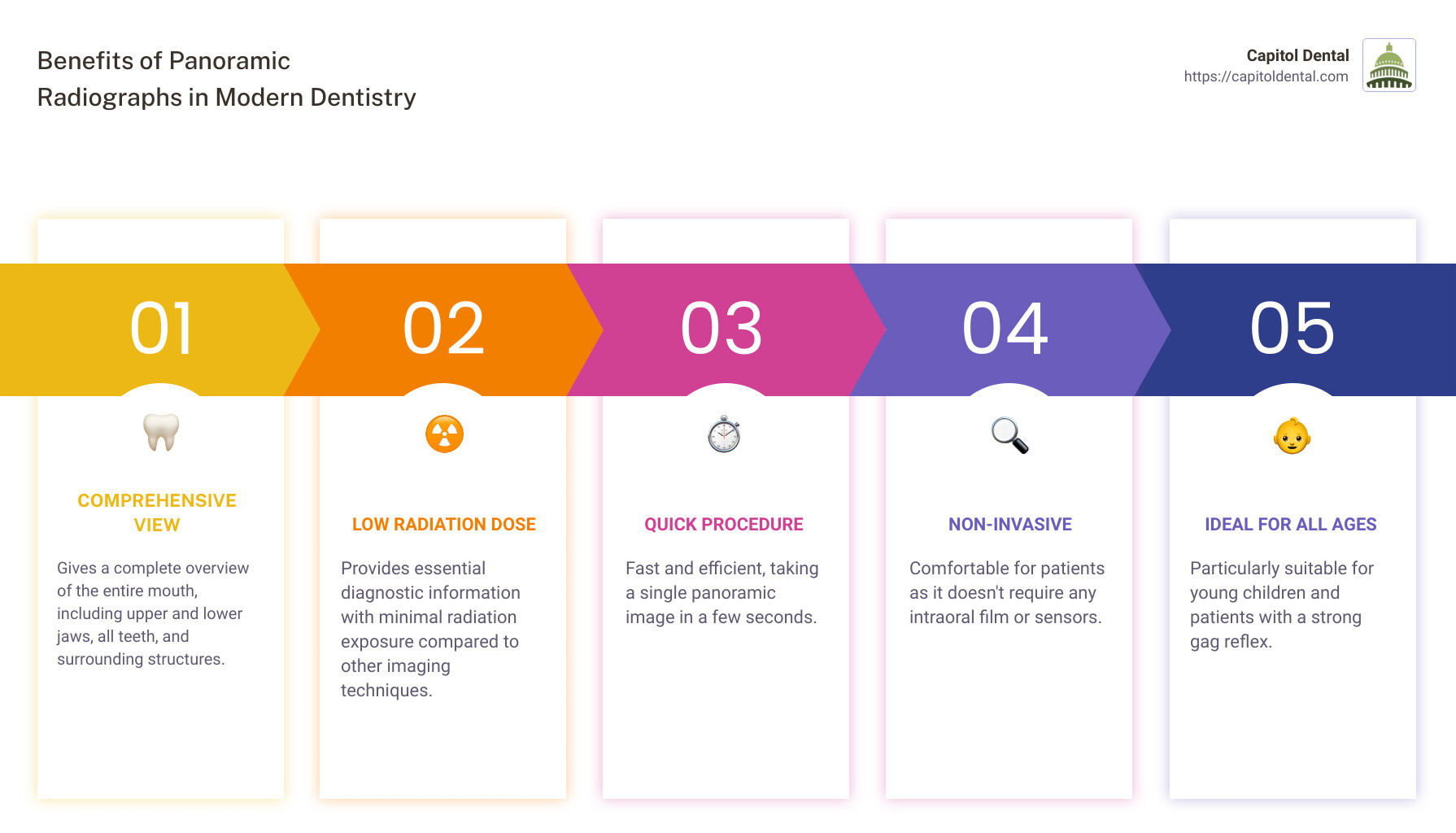
Comprehensive View
A dental panoramic radiograph captures the entire mouth in a single image. This includes all teeth, the upper and lower jaws, and surrounding structures. This broad view helps dentists spot issues that might be missed with traditional x-rays. For example, they can see impacted teeth, jaw tumors, and even sinus problems.
Low Radiation Dose
One of the top concerns with any x-ray is radiation exposure. The good news is that panoramic radiographs use a very small dose of ionizing radiation. Research shows that the radiation from a panoramic x-ray is comparable to just a few days of natural background radiation. This makes it a safer option, especially for young children and pregnant women.
Quick Procedure
The process of taking a panoramic radiograph is quick and straightforward. It usually takes about 18 seconds for the machine to capture the image. This speed is beneficial for both the patient and the dentist, allowing for more efficient appointments.
Non-Invasive
Unlike traditional x-rays that require placing film or sensors inside the mouth, panoramic x-rays are non-invasive. The x-ray tube and detector rotate around the outside of your head. This is particularly helpful for patients with a strong gag reflex or those who find it uncomfortable to have objects placed in their mouth.
Ideal for Young Children
For young children, traditional x-rays can be challenging. They may not sit still, or they might be scared of the equipment. Panoramic radiographs are less intimidating and quicker, making it easier to get accurate images without causing distress.
By offering a comprehensive view, using a low radiation dose, and being a quick, non-invasive procedure, panoramic radiographs are a valuable tool in modern dentistry. Next, let’s explore some of the limitations of panoramic radiographs.
Limitations of Panoramic Radiographs
Distortions and Magnifications
While panoramic radiographs provide a broad view of the mouth, they can sometimes produce distortions and magnifications. Because the image is a two-dimensional representation of a three-dimensional structure, certain areas might appear stretched or compressed. This can make it difficult to get precise measurements or see small details clearly.
Blurry Images
Another limitation is that areas outside the focal path can become blurry. The focal path is the specific area where the anatomical features are captured sharply. Anything outside this path might not be as clear, which can lead to missed details or misinterpretations.
Detailed Information
For cases requiring detailed information, such as diagnosing specific tooth issues or soft tissue conditions, panoramic radiographs might not be sufficient. They don’t provide the level of detail necessary for certain diagnoses and treatments.
CT Scan and MRI
In situations where more detailed imaging is needed, dentists may recommend a CT scan or MRI. These imaging techniques offer more precise and comprehensive views of the teeth, bones, and soft tissues. For example, Dental Cone Beam CT is specifically designed for detailed imaging of the dental structures and is often used for implant planning and complex diagnoses.
While panoramic radiographs are a useful tool, understanding their limitations helps ensure that patients receive the most accurate and effective care.
Next, let’s discuss the safety and preparation for panoramic radiographs.
Safety and Preparation for Panoramic Radiographs
Radiation Exposure
Panoramic dental x-rays use a very small dose of ionizing radiation to produce images of your entire mouth. The exposure is minimal, significantly lower than traditional full-mouth periapical radiography. According to a review, the risk of fatal cancer from a panoramic radiograph is about 1 in 20,000,000. This low risk makes the procedure safe for routine dental examinations.
Lead Apron
Historically, patients were provided with a lead apron to protect their body from radiation exposure. However, recent guidelines from the American Dental Association (ADA) suggest that lead aprons may no longer be necessary due to advancements in X-ray technology. These advancements have made the radiation exposure even lower and more targeted.
Pregnancy Precautions
If there’s a possibility you might be pregnant, inform your dentist. While the radiation dose from a panoramic x-ray is very low, precautionary measures are often taken to minimize any risk to the fetus. In some cases, your dentist may decide to delay the x-ray until after the pregnancy unless it’s absolutely necessary.
Remove Metal Objects
Before undergoing a panoramic radiograph, you’ll need to remove any metal objects from the region being imaged. This includes:
- Jewelry
- Eyeglasses
- Removable orthodontic appliances
- Dentures
- Hairpins
Removing these items helps prevent artifacts on the image, ensuring a clear and accurate radiograph.
By understanding these safety measures and preparation steps, you can ensure a smooth and worry-free experience during your panoramic dental x-ray.
Next, let’s address some frequently asked questions about panoramic radiographs.
Frequently Asked Questions about Panoramic Radiographs
What can panoramic radiographs detect?
A dental panoramic radiograph can detect a wide range of dental and oral health issues. Here are some key things it can identify:
- Dental Caries: While not as effective for small cavities, panoramic radiographs can spot larger carious lesions.
- Periodontal Diseases: These x-rays can reveal bone loss around teeth, indicating gum disease.
- Lesions and Tumors: Dentists can identify cysts, tumors, and other abnormalities in the jaws and surrounding structures.
- Impacted Teeth: Wisdom teeth and other impacted teeth are clearly visible.
- Jaw Disorders: Problems with the temporomandibular joint (TMJ) can be assessed.
- Sinus Issues: Since the sinus cavities are included, sinusitis and other sinus problems can be detected.
What are the disadvantages of panoramic radiographs?
While panoramic radiographs are incredibly useful, they do have some limitations:
- Distortions and Magnifications: The curved nature of the mouth can cause slight distortions and magnifications, making some areas appear larger or smaller than they are.
- Blurry Images: Sometimes, the image can be slightly blurry, especially if the patient moves during the scan.
- Lesion Identification: Small lesions and cavities may not be as easily detected compared to intraoral x-rays.
How often should panoramic radiographs be taken?
The frequency of taking panoramic radiographs depends on individual needs and dental health:
- Routine Check-Ups: For most people, a panoramic radiograph is recommended every five to eight years.
- Treatment Planning: If you are undergoing specific treatments like braces, implants, or extractions, your dentist might recommend more frequent scans.
- Dental Check-Ups: Your dentist will decide based on your dental history and current health. Regular check-ups help in early detection and better treatment planning.
Panoramic radiographs are a vital tool in modern dentistry, offering a comprehensive view of the mouth with minimal discomfort and radiation exposure. Understanding what they can and cannot do helps in making informed decisions about your dental care.
Next, let’s wrap up with some concluding thoughts on the benefits of choosing Capitol Dental for your panoramic radiograph and other dental needs.
Conclusion
At Capitol Dental, we pride ourselves on providing comprehensive dental services using the latest advancements in technology. Our use of panoramic radiographs exemplifies our commitment to offering top-notch care that is both thorough and efficient.
Advanced Technology is at the core of our practice. By utilizing digital panoramic radiographs, we can quickly and accurately diagnose a wide range of dental issues, from cavities and periodontal diseases to more complex conditions like jaw tumors and TMJ disorders. This allows us to create detailed and personalized treatment plans tailored to each patient’s needs.
One of the standout benefits of panoramic radiographs is their ability to give a complete view of your mouth in just a single, quick scan. This means less time in the chair and more accurate diagnoses, helping to ensure that no potential issues are overlooked. Plus, the low radiation dose and non-invasive nature make it a safe option for patients of all ages, including young children and those with a sensitive gag reflex.
At Capitol Dental, we understand that each patient is unique. That’s why we emphasize personalized care. Our skilled team takes the time to listen to your concerns, explain your options, and develop a treatment plan that fits your specific needs and preferences. We believe that informed patients are empowered patients, and we strive to make your dental experience as comfortable and stress-free as possible.
Ready to experience the benefits of advanced dental care? Contact Capitol Dental today to schedule your appointment and take the first step toward a healthier, brighter smile.
By choosing Capitol Dental, you are opting for a practice that values your health, comfort, and satisfaction. We look forward to helping you achieve your best oral health.